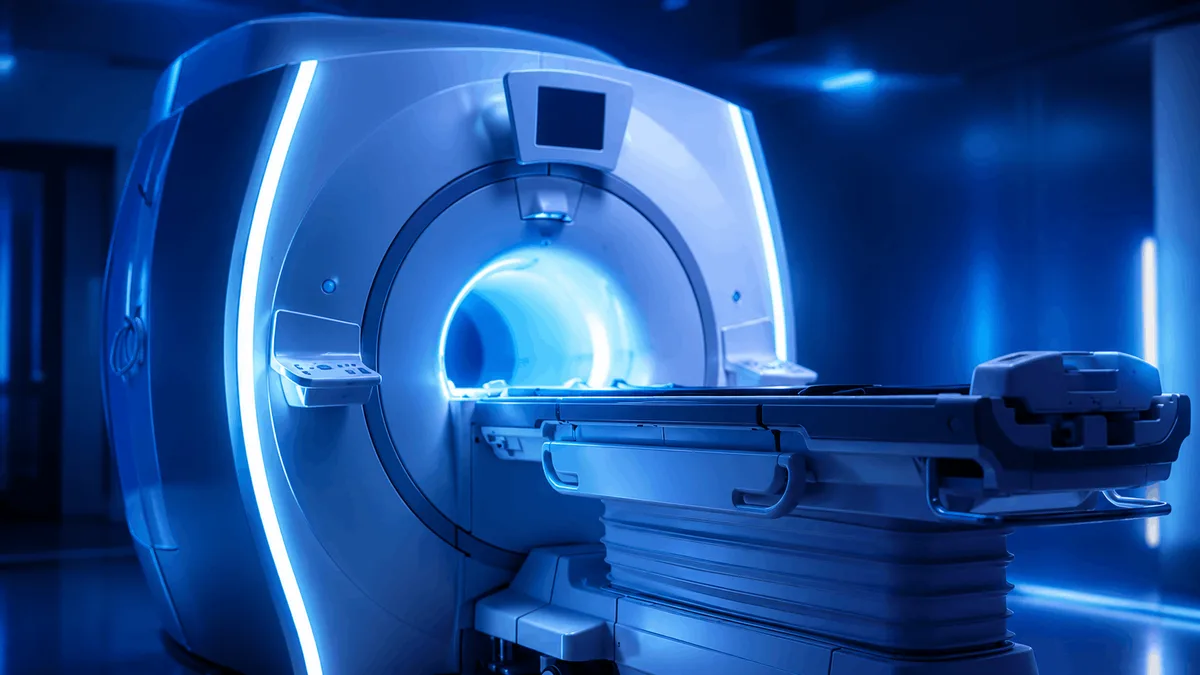
PET MRI scan
PET/MRI is an advanced hybrid imaging test that combines positron emission tomography (PET) with magnetic resonance imaging (MRI) in a single examination. It provides detailed information about both the metabolic activity of tissues and their anatomical structure, making it particularly useful in selected cancer, neurological and complex soft-tissue conditions.
Quick Summary
PET MRI scan is offered by 1 vetted provider across 1 destination in the Macrocare network. Prices start from $1,050, excluding flights and accommodation.
What is a PET/MRI scan?
A PET/MRI scan, also known as positron emission tomography with magnetic resonance imaging, is an advanced diagnostic imaging examination that combines two different imaging technologies in a single system.
PET (Positron Emission Tomography) provides information about the metabolic, molecular and biological activity of tissues.
MRI (Magnetic Resonance Imaging) produces highly detailed images of organs, soft tissues, the brain, bone marrow and other structures using a strong magnetic field and radiofrequency waves.
By combining both techniques, PET/MRI can show not only where an abnormality is located, but also how biologically active it is.
Before the PET part of the examination, a small amount of radioactive material called a radiotracer or radiopharmaceutical is administered. One of the most commonly used tracers is 18F-FDG (fluorodeoxyglucose), a glucose-like compound that can accumulate in tissues with increased metabolic activity.
Other PET tracers may be used depending on the disease being investigated and the clinical purpose of the scan.
PET/MRI may help physicians diagnose disease, assess its extent, plan treatment, evaluate treatment response and investigate suspected recurrence.
How does PET/MRI work?
PET and MRI provide complementary types of information.
PET component
A small amount of radiotracer is usually injected into a vein.
The tracer travels through the body and accumulates in different tissues according to their biological activity.
For example, FDG behaves similarly to glucose. Some tissues with increased glucose metabolism may absorb more FDG and therefore appear more active on PET images.
Increased radiotracer uptake does not always indicate cancer. Infection, inflammation and some normal tissues may also show increased uptake.
MRI component
MRI uses:
- A strong magnetic field
- Radiofrequency pulses
- Specialized receiver coils
- Computer processing
to create detailed images of the body's internal structures.
MRI is particularly useful for evaluating:
- Brain and spinal tissues
- Liver
- Pelvic organs
- Muscles
- Bone marrow
- Soft-tissue tumors
- Blood vessels
- Other structures requiring high soft-tissue contrast
Combined imaging
PET and MRI data are combined so that areas of abnormal molecular activity can be precisely correlated with anatomical structures.
This may improve localization and characterization of disease in appropriately selected patients.
What is PET/MRI used for?
PET/MRI has applications in several specialties, particularly oncology, neurology and selected complex diagnostic conditions.
PET/MRI in cancer
PET/MRI may be used in selected patients to:
- Detect and characterize certain tumors
- Determine the location and extent of cancer
- Assist with cancer staging
- Evaluate lymph nodes
- Assess possible metastatic spread
- Evaluate bone and bone marrow involvement
- Examine complex soft-tissue tumors
- Help plan surgery
- Help plan radiotherapy
- Evaluate response to chemotherapy
- Evaluate response to immunotherapy
- Evaluate response to targeted therapy
- Investigate suspected residual disease
- Evaluate suspected cancer recurrence
- Compare findings with previous imaging
The value of PET/MRI depends on the tumor type, the radiotracer used and the clinical question.
Cancers where PET/MRI may be considered
Depending on specialist assessment and local protocols, PET/MRI may be considered in selected cases involving:
- Brain tumors
- Head and neck cancers
- Lymphoma
- Breast cancer
- Prostate cancer
- Gynecological cancers
- Cervical cancer
- Ovarian cancer
- Rectal cancer
- Liver tumors
- Pancreatic tumors
- Bone tumors
- Soft-tissue sarcomas
- Multiple myeloma
- Pediatric cancers
- Metastatic cancer
- Tumors requiring detailed soft-tissue or bone marrow assessment
Not every patient with cancer requires PET/MRI. The appropriate test may instead be PET/CT, MRI, CT or another imaging examination depending on the case.
PET/MRI for brain and neurological conditions
PET/MRI can combine detailed structural brain imaging with molecular information from PET.
Potential applications in selected patients include:
- Evaluation of certain brain tumors
- Assessment of suspected brain tumor recurrence
- Treatment planning for brain tumors
- Evaluation of epilepsy before surgery in selected cases
- Assessment of certain neurodegenerative disorders
- Specialized evaluation of cognitive impairment or dementia using appropriate PET tracers
- Assessment of brain metabolism and molecular processes in selected clinical situations
The choice of PET radiotracer depends on the indication.
PET/MRI in children and young patients
PET/MRI may be particularly useful in selected children, adolescents and young adults who require molecular imaging.
Because the MRI component does not use X-rays, PET/MRI may reduce cumulative ionizing radiation exposure compared with PET/CT in some protocols.
This can be an important consideration in younger patients who may require repeated imaging.
However, PET/MRI can take longer than many conventional scans, and some young children may require sedation or anesthesia if they are unable to remain still.
PET/MRI vs PET/CT
PET/MRI and PET/CT both combine PET molecular imaging with an anatomical imaging technique.
PET/MRI combines:
- PET molecular and metabolic imaging
- High-resolution MRI
- Excellent soft-tissue contrast
- Functional MRI techniques when clinically required
PET/CT combines:
- PET molecular imaging
- Computed tomography using X-rays
A potential advantage of PET/MRI is that the MRI component does not use ionizing radiation.
The patient is still exposed to a small amount of radiation from the PET radiotracer, but total radiation exposure may be lower than with comparable PET/CT examinations.
PET/MRI can be particularly valuable when detailed soft-tissue evaluation is important.
However, PET/MRI is not automatically better than PET/CT. The most appropriate examination depends on the disease, body area, clinical question and required imaging information.
What happens during a PET/MRI scan?
The exact procedure depends on the radiotracer and the type of examination.
A typical PET/MRI appointment may include the following steps.
Medical assessment
Before the scan, the medical team may review:
- The reason for the examination
- Medical history
- Previous imaging
- Previous cancer treatment
- Current medications
- Diabetes
- Kidney disease
- Pregnancy possibility
- Breastfeeding
- Allergies
- Previous reactions to MRI contrast
- Implanted medical devices
- Metal inside the body
Blood glucose measurement
For some FDG PET examinations, blood glucose may be checked before radiotracer administration.
Blood glucose levels can affect FDG distribution and image quality.
Patients with diabetes usually receive specific preparation instructions.
Radiotracer injection
A small intravenous line is inserted, usually into the arm or hand.
The PET radiotracer is then administered.
Uptake period
After administration of certain radiotracers such as FDG, the patient usually rests quietly while the tracer distributes through the body.
This uptake period is commonly approximately 30 to 60 minutes, although timing varies according to the tracer and imaging protocol.
Patients may be asked to:
- Rest quietly
- Avoid unnecessary movement
- Avoid excessive talking
- Remain warm and comfortable
PET/MRI imaging
The patient lies on a padded examination table that moves into the PET/MRI scanner.
Special MRI coils may be positioned around the area being examined.
During MRI imaging, the scanner produces loud tapping, knocking or buzzing sounds. Hearing protection is usually provided.
The patient must remain as still as possible because movement can reduce image quality.
The technologist can communicate with the patient during the examination.
How long does PET/MRI take?
PET/MRI examinations are often longer than routine CT or MRI scans.
The total appointment may take approximately 1.5 to 3 hours or longer, depending on:
- The radiotracer
- Uptake time
- Area of the body being examined
- Whole-body or focused imaging
- Number of MRI sequences
- Whether MRI contrast is required
- Need for delayed or additional images
- Patient movement
- Need for sedation or anesthesia
The actual scanning period is only part of the total appointment time.
How should I prepare for PET/MRI?
Preparation varies according to the radiotracer and examination protocol.
For many FDG PET examinations, patients may be instructed to:
- Avoid food for several hours before the scan
- Drink plain water
- Avoid sugary or calorie-containing drinks during the fasting period
- Follow specific instructions regarding medications
- Follow special instructions if diabetic
- Avoid strenuous physical activity before the scan when advised
- Arrive at the imaging center at the scheduled time
Patients should follow the preparation instructions provided by the imaging center carefully because incorrect preparation can affect PET image quality.
What should I tell the medical team before PET/MRI?
Inform the imaging center if you:
- Are pregnant or may be pregnant
- Are breastfeeding
- Have diabetes
- Have kidney disease
- Have reduced kidney function
- Have severe liver disease
- Have allergies
- Have had a previous reaction to gadolinium MRI contrast
- Have severe claustrophobia
- Have difficulty remaining still
- Have an implanted medical device
- Have a pacemaker
- Have an implantable defibrillator
- Have a cochlear implant
- Have an implanted neurostimulator
- Have an insulin or medication pump
- Have aneurysm clips
- Have metallic fragments, bullets or shrapnel in the body
- May have metallic fragments in the eyes
- Have undergone surgery involving implanted devices
Many modern implants are MRI-compatible or MR Conditional, but the exact device model and scanning conditions must be checked before MRI.
Whenever possible, bring the implant identification card or device information.
MRI safety
MRI uses a powerful magnetic field.
Metallic and electronic objects must be removed or assessed before entering the scanner room.
These may include:
- Jewelry
- Watches
- Mobile phones
- Hearing aids
- Removable dental devices
- Keys
- Coins
- Credit cards
- Clothing containing metallic components
- Other metallic items
Some implanted devices require specialist assessment before MRI.
PET/MRI should only be performed after appropriate MRI safety screening.
Will MRI contrast be used?
Not every PET/MRI scan requires intravenous MRI contrast.
When additional anatomical detail is needed, a gadolinium-based contrast agent may be administered.
Inform the medical team if you have:
- Significant kidney disease
- Kidney failure
- Kidney transplant
- Previous reaction to gadolinium
- Pregnancy or possible pregnancy
Kidney function may need to be assessed before contrast administration in selected patients.
Is PET/MRI painful?
PET/MRI is generally noninvasive and usually painless apart from intravenous cannula insertion and injection.
Some patients may experience discomfort from:
- Remaining still for a long period
- Lying inside the MRI scanner
- Loud scanner sounds
- Claustrophobia
- Intravenous cannula insertion
Patients with significant anxiety or claustrophobia should inform the imaging center before the examination.
Is PET/MRI safe?
PET/MRI is generally considered safe when performed for an appropriate clinical indication and after proper screening.
The PET component uses a small amount of radioactive material.
The MRI component does not use ionizing radiation.
Radiotracers naturally lose radioactivity over time and are eliminated from the body.
Patients are often encouraged to drink fluids after the scan, unless medically restricted, to help eliminate the radiotracer through the urine.
PET/MRI and pregnancy
Because PET uses radioactive material, pregnancy requires special consideration.
Patients who are pregnant or could be pregnant should inform their doctor and the nuclear medicine team before radiotracer administration.
The medical team will determine whether:
- The examination should proceed
- It should be postponed
- The protocol should be modified
- Another imaging method would be more appropriate
Gadolinium MRI contrast is also generally avoided during pregnancy unless considered clinically necessary.
PET/MRI and breastfeeding
Patients who are breastfeeding should inform the medical team before the examination.
Breastfeeding recommendations depend on the specific PET radiotracer used.
Temporary interruption may be recommended with some radiopharmaceuticals.
The nuclear medicine department should provide tracer-specific instructions.
PET/MRI in patients with diabetes
Diabetes can affect FDG PET imaging because blood glucose levels influence FDG distribution.
Patients with diabetes may receive individualized instructions regarding:
- Fasting
- Meal timing
- Insulin
- Oral diabetes medications
- Appointment timing
- Blood glucose targets
Do not stop insulin or diabetes medication unless specifically instructed by the medical team.
What happens after PET/MRI?
Unless sedation has been used, most patients can usually leave the imaging center shortly after the scan.
Patients can generally return to normal activities unless advised otherwise.
The medical team may recommend:
- Drinking adequate fluids
- Emptying the bladder regularly
- Following tracer-specific radiation precautions
- Following breastfeeding instructions when applicable
- Following post-sedation instructions if sedation was used
Who interprets a PET/MRI scan?
PET/MRI requires specialized interpretation.
Depending on the healthcare facility and the type of examination, images may be reviewed by physicians experienced in:
- Nuclear medicine
- Diagnostic radiology
- MRI
- Oncological imaging
- Neuroradiology
- Other relevant subspecialties
PET and MRI findings are interpreted together with the patient's medical history and previous examinations.
What does a PET/MRI report show?
A PET/MRI report may describe:
- Areas of abnormal radiotracer uptake
- Intensity and pattern of uptake
- Anatomical abnormalities on MRI
- Tumor size and local extent
- Lymph-node findings
- Possible metastatic disease
- Bone or bone marrow abnormalities
- Comparison with previous imaging
- Treatment response
- Findings requiring further investigation
PET findings must always be interpreted in clinical context.
Increased tracer uptake does not necessarily mean cancer because infection, inflammation and normal physiological activity may also produce increased uptake.
Some tumors may also show relatively low uptake with certain radiotracers.
What does SUV mean in PET imaging?
Some PET reports include a measurement called the Standardized Uptake Value (SUV).
SUV is a semi-quantitative measurement of radiotracer uptake in a particular area.
It can help physicians assess areas of activity or compare changes over time.
However, SUV cannot diagnose cancer by itself.
SUV values must be interpreted together with:
- MRI findings
- Pattern of tracer uptake
- Tumor type
- Patient history
- Treatment history
- Previous imaging
Benefits of PET/MRI
Potential benefits include:
- Molecular and anatomical imaging in a single examination
- Excellent soft-tissue visualization
- Precise correlation of PET abnormalities with MRI anatomy
- Lower ionizing radiation exposure than PET/CT in many protocols
- High-quality brain imaging
- Detailed pelvic imaging
- Detailed liver imaging
- Soft-tissue tumor assessment
- Bone marrow evaluation
- Potential advantages in children and younger patients
- Evaluation of treatment response
- Potential reduction in the need for separate PET and MRI appointments in selected patients
Limitations of PET/MRI
PET/MRI also has limitations, including:
- Limited availability compared with PET/CT
- Longer examination time
- Higher cost in some healthcare systems
- Need to remain still for an extended period
- Claustrophobia
- MRI restrictions for some implants or metallic devices
- Increased PET uptake may occur in inflammation and infection
- Some diseases or anatomical structures may be better evaluated with CT
- Not every disease requires PET/MRI
- Diagnostic value depends on choosing the correct radiotracer and imaging protocol
The referring physician and imaging specialist should determine whether PET/MRI is appropriate for each patient.
Can PET/MRI detect cancer early?
PET can identify abnormal metabolic or molecular activity and may detect disease before major anatomical changes become visible in some situations.
However, PET/MRI is not a universal cancer screening test.
Its accuracy varies according to:
- Cancer type
- Tumor biology
- Lesion size
- Radiotracer used
- Blood glucose level
- Previous treatment
- Imaging protocol
A normal PET/MRI does not completely exclude cancer, and an abnormal scan does not automatically confirm cancer.
Can PET/MRI determine whether a tumor is benign or malignant?
PET/MRI can provide important information about a lesion, but imaging alone cannot always determine whether a tumor is benign or malignant.
Additional assessment may include:
- Clinical examination
- Laboratory tests
- Follow-up imaging
- Endoscopy
- Biopsy
- Histopathological examination
A biopsy may still be required to establish a definitive diagnosis.
Can PET/MRI assess response to cancer treatment?
Yes. In selected patients, PET/MRI may help evaluate treatment response.
PET can demonstrate changes in tumor metabolic activity, while MRI can assess changes in tumor size, anatomy and tissue characteristics.
PET/MRI may be used to evaluate response to:
- Chemotherapy
- Immunotherapy
- Targeted therapy
- Radiotherapy
- Other systemic cancer treatments
The appropriate timing of the scan depends on the cancer type and treatment.
Can PET/MRI detect cancer recurrence?
PET/MRI may help investigate suspected recurrent cancer in selected patients, especially when both molecular information and detailed soft-tissue imaging are important.
However, inflammation and healing after treatment can sometimes cause increased PET activity, so the timing of the examination and specialist interpretation are important.
Diagnosis and Treatment
PET/MRI is not a treatment. It is an advanced diagnostic imaging test that may influence several stages of patient management.
Determining disease extent
PET/MRI may help identify the primary abnormality, surrounding tissue involvement, lymph-node disease and possible distant spread.
Cancer staging
Accurate staging can influence whether treatment involves surgery, radiotherapy, systemic therapy or a combination of treatments.
Treatment selection
Molecular and anatomical information may help multidisciplinary teams determine the most appropriate treatment strategy.
Surgical planning
MRI provides detailed information about the relationship between a tumor and surrounding structures, while PET can provide information about biological activity.
Radiotherapy planning
Hybrid imaging may help physicians identify active disease and define treatment targets in selected cases.
Evaluating treatment response
PET may show changes in metabolic activity, while MRI can demonstrate changes in size, anatomy and tissue characteristics.
Investigating suspected recurrence
PET/MRI may help identify suspicious abnormalities after treatment and determine whether further investigation is required.
Guiding additional tests or biopsy
When an abnormal area is identified, PET/MRI findings may help physicians decide whether targeted biopsy or additional imaging is necessary.
Final treatment decisions should always be based on the complete clinical picture, including pathology, laboratory results, imaging and specialist assessment.
Frequently Asked Questions
Is PET/MRI the same as PET/CT?
No. PET/MRI combines PET with magnetic resonance imaging, while PET/CT combines PET with computed tomography.
Does PET/MRI use radiation?
Yes. The PET radiotracer involves a small amount of ionizing radiation. The MRI component itself does not use ionizing radiation.
Do I need to fast before PET/MRI?
Fasting is commonly required for FDG PET imaging, but preparation depends on the radiotracer and protocol. Follow the instructions provided by the imaging center.
Can I drink water before the scan?
Plain water is commonly allowed and may be encouraged for many FDG PET examinations unless the medical team provides different instructions.
Can patients with diabetes undergo PET/MRI?
Yes. Special preparation may be required to optimize blood glucose levels and image quality.
Can I take my medications before the scan?
Many medicines can be continued, but instructions vary according to the examination. Inform the imaging center about all medications and supplements.
Can I undergo PET/MRI if I have a pacemaker?
Some modern pacemakers and implanted devices are MR Conditional and can be scanned under specific conditions. The exact device must be assessed before the examination.
Is PET/MRI suitable for children?
It may be useful in selected pediatric cases, particularly because MRI does not use X-rays and may help reduce cumulative radiation exposure compared with PET/CT.
Will I need sedation?
Most adults do not require sedation. It may be considered for severe claustrophobia or for young children who cannot remain still.
How soon will I receive the results?
Reporting time varies between healthcare facilities and according to case complexity. PET/MRI often requires detailed review of both PET and MRI images and comparison with previous studies.
SEO Title
PET/MRI Scan: Uses, Preparation, Procedure & Results
Meta Description
Learn about PET/MRI scans, including cancer and brain imaging uses, preparation, procedure, radiation, safety, results and the difference between PET/MRI and PET/CT.
Frequently Asked Questions
PET MRI scan starts from $1,050 through the Macrocare network, available in Cairo. The exact price depends on the provider, the technique used and your medical assessment — request a free quotation for a confirmed figure.
PET MRI scan is offered by 1 verified providers in the Macrocare network across Cairo. Each listing shows the provider, the city and its real starting price.
Send a free quotation request and a Macrocare coordinator compares verified providers for PET MRI scan, confirms pricing, and arranges appointments, travel and follow-up. Coordination is free for patients.
PET MRI scan belongs to Advanced Diagnostics. Browse that category to compare related options, their verified providers and real starting prices before deciding.
Available in
Related suggestions
More in Advanced Diagnostics
Compare related options with their verified providers and real starting prices.
MRI Scan
27 verified providers
Biopsy Analysis
5 verified providers
Colonoscopy
5 verified providers
Full-Body Health Checkup
4 verified providers
Ultrasound Imaging
3 verified providers
Bone Density Scan ( DEXA )
2 verified providers
Listed prices only — travel and stay are estimated separately. Final cost confirmed on your free quotation. Rates as of 2026-07-20.