Dr. Ahmed Sameh Nada – Consultant Neurosurgeon, Neurointervention and Spine Surgery Specialist
Dr. Ahmed Sameh Nada is a Consultant and Lecturer of Neurosurgery, specializing in brain surgery, spine surgery, cerebrovascular neurosurgery, and neurointerventional procedures.
Dr. Ahmed Sameh Nada combines conventional microsurgical neurosurgery with modern Neurointervention, allowing complex neurological and vascular conditions to be assessed from both surgical and endovascular perspectives.
His expertise includes brain aneurysms, cerebral arteriovenous malformations, AVM, dural arteriovenous fistulas, DAVF, acute ischemic stroke thrombectomy, carotid and intracranial artery stenosis, brain and skull base tumors, cervical and lumbar disc disease, spinal stenosis, and spinal fixation surgery.
Through Macrocare, patients can submit their medical reports, MRI, CT, angiography, and previous procedure images for case review and appointment coordination with Dr. Ahmed Sameh Nada.
Dr. Ahmed Sameh Nada’s Specialties
The expertise of Dr. Ahmed Sameh Nada in neurosurgery and neurointervention includes:
- Diagnostic cerebral angiography.
- Therapeutic cerebral angiography.
- Brain aneurysm treatment.
- Brain aneurysm coiling.
- Balloon-assisted coiling.
- Stent-assisted coiling.
- Flow diverter stents.
- Advanced aneurysm occlusion devices.
- Microsurgical clipping of brain aneurysms.
- Treatment of giant and complex cerebral aneurysms.
- Mechanical thrombectomy for acute ischemic stroke.
- Stent retriever thrombectomy.
- Aspiration thrombectomy.
- Treatment of Middle Cerebral Artery occlusion.
- Treatment of Basilar Artery occlusion.
- Brain AVM treatment.
- Brain AVM embolization.
- Dural arteriovenous fistula treatment.
- Carotid cavernous fistula treatment.
- Spinal vascular malformation treatment.
- Carotid artery stenosis treatment.
- Carotid artery stenting.
- Intracranial artery stenosis evaluation.
- Brain tumor surgery.
- Skull base tumor surgery.
- Meningioma surgery.
- Pituitary tumor surgery.
- Glioma surgery.
- Acoustic neuroma surgery.
- Lumbar disc surgery.
- Cervical disc surgery.
- Spinal stenosis surgery.
- Spinal fixation.
- Spondylolisthesis treatment.
- Spinal fracture surgery.
- Minimally invasive spine surgery.
- Radiofrequency treatment for selected spinal pain conditions.
- Traumatic brain injury surgery.
- Acute intracranial hemorrhage management.
- Epidural and subdural hematoma surgery.
- Hydrocephalus treatment.
- VP shunt surgery.
Cerebral Angiography and Neurointervention with Dr. Ahmed Sameh Nada
Cerebral Angiography and Neurointervention are used to diagnose and treat vascular diseases affecting the brain and spinal cord.
These techniques may be used to assess:
- Brain aneurysms.
- Arteriovenous malformations.
- Dural arteriovenous fistulas.
- Carotid cavernous fistulas.
- Arterial narrowing or occlusion.
- Selected spinal vascular abnormalities.
In some cases, diagnosis and treatment can be performed through the same endovascular pathway.
What Is Cerebral Angiography?
Cerebral angiography is a catheter-based procedure used to visualize blood vessels supplying the brain.
A thin catheter is introduced through an artery, commonly from the groin or wrist, and guided toward the cerebral circulation.
The procedure may be used for:
- Diagnostic cerebral angiography.
- Aneurysm coiling.
- Embolization.
- Stent placement.
- Mechanical thrombectomy.
- Flow diverter placement.
The exact technique depends on the diagnosis.
Brain Aneurysm Treatment with Dr. Ahmed Sameh Nada
A Brain Aneurysm is a weakened, bulging area in the wall of a cerebral artery.
Aneurysms may be discovered:
- Incidentally on imaging.
- During evaluation of headaches or neurological symptoms.
- After subarachnoid hemorrhage.
- During assessment of another neurological condition.
Treatment depends on aneurysm size, location, shape, rupture status, patient age, and other risk factors.
Does Every Brain Aneurysm Need Treatment?
No.
Some small aneurysms may be monitored with imaging, while others require treatment because of their size, anatomy, symptoms, or rupture risk.
Assessment may include:
- Aneurysm size.
- Location.
- Shape and neck width.
- Presence of symptoms.
- Previous rupture.
- Family history.
- Patient age.
- Associated risk factors.
Dr. Ahmed Sameh Nada evaluates whether observation, endovascular treatment, or microsurgical treatment is more appropriate.
Brain Aneurysm Coiling
Aneurysm Coiling is an endovascular technique used to treat selected cerebral aneurysms.
Small coils are placed inside the aneurysm through a catheter to reduce blood flow into it and promote isolation of the aneurysm from the circulation.
Suitability depends on aneurysm anatomy and location.
Balloon-Assisted Coiling
Balloon-Assisted Coiling may be used for selected aneurysms with a broad neck.
A temporary balloon is positioned across the aneurysm neck while coils are inserted to help keep them within the aneurysm.
Stent-Assisted Coiling
Stent-Assisted Coiling may be used for wide-neck cerebral aneurysms.
A stent is placed inside the parent artery to provide support while coils are inserted into the aneurysm.
Flow Diverter Treatment
Flow Diverter Stents are specialized devices used to treat selected cerebral aneurysms.
They redirect blood flow along the normal artery and reduce flow into the aneurysm, allowing gradual thrombosis and vessel healing.
Flow diversion may be considered for selected:
- Large aneurysms.
- Wide-neck aneurysms.
- Complex aneurysms.
- Aneurysms that are difficult to treat with conventional coiling.
Microsurgical Clipping of Brain Aneurysms
In some patients, Microsurgical Clipping is more appropriate than endovascular treatment.
During surgery, a clip is placed across the aneurysm neck to exclude it from the circulation.
The choice between clipping and coiling depends on aneurysm anatomy, location, rupture status, patient age, and other clinical factors.
Subarachnoid Hemorrhage from a Ruptured Brain Aneurysm
A ruptured aneurysm can cause Subarachnoid Hemorrhage, SAH.
Symptoms may include:
- Sudden severe headache.
- Vomiting.
- Loss of consciousness.
- Neck stiffness.
- Seizures.
- Neurological weakness.
This is a medical emergency requiring urgent assessment in a specialized center.
Mechanical Thrombectomy with Dr. Ahmed Sameh Nada
Mechanical Thrombectomy is a catheter-based procedure used to remove certain large blood clots causing acute ischemic stroke.
Time is a critical factor in stroke care.
In suitable patients, a catheter is advanced to the blocked artery and the clot is removed to restore cerebral blood flow.
When Is Mechanical Thrombectomy Used?
Patient selection depends on factors such as:
- Time of symptom onset.
- Location of the blocked artery.
- Extent of established brain injury.
- Amount of potentially salvageable brain tissue.
- CT Brain findings.
- CT Angiography.
- CT Perfusion in selected patients.
- MRI or MR Perfusion in selected situations.
Not every ischemic stroke is suitable for thrombectomy.
Symptoms of Acute Stroke
Urgent symptoms include:
- Sudden facial weakness.
- Sudden weakness of an arm or leg.
- Sudden difficulty speaking.
- Sudden inability to understand speech.
- Sudden visual disturbance.
- Sudden loss of balance.
- Severe unexplained dizziness.
- Reduced consciousness.
Stroke is an emergency and requires immediate hospital evaluation.
Stent Retriever Thrombectomy
A Stent Retriever can be deployed inside the blocked artery to engage the clot and remove it.
The technique is selected according to clot location, anatomy, and procedural strategy.
Aspiration Thrombectomy
Aspiration Thrombectomy uses a specialized catheter to apply suction directly to the clot.
It may be used alone or in combination with other thrombectomy techniques.
Middle Cerebral Artery Occlusion
Middle Cerebral Artery, MCA, Occlusion is a common cause of large-vessel ischemic stroke.
It may cause:
- Severe weakness on one side.
- Facial drooping.
- Speech difficulty.
- Sensory loss.
- Reduced consciousness in severe cases.
Selected patients may benefit from mechanical thrombectomy.
Basilar Artery Occlusion
Basilar Artery Occlusion is a serious posterior circulation stroke.
Possible symptoms include:
- Reduced consciousness.
- Weakness of multiple limbs.
- Severe balance disturbance.
- Double vision.
- Difficulty speaking or swallowing.
- Coma in severe cases.
Urgent imaging and neurovascular assessment are required to determine whether endovascular treatment is appropriate.
Brain AVM Treatment with Dr. Ahmed Sameh Nada
A Brain Arteriovenous Malformation, AVM, is an abnormal connection between arteries and veins in the brain.
It may present with:
- Intracranial hemorrhage.
- Seizures.
- Headache.
- Weakness or numbness.
- Other neurological symptoms.
Treatment depends on AVM size, location, vascular anatomy, and rupture risk.
Brain AVM Embolization
Endovascular Embolization can be used to close selected blood vessels feeding an AVM.
It may be used:
- As a primary treatment in selected cases.
- Before microsurgery.
- Before stereotactic radiosurgery.
- As part of a combined treatment strategy.
Microsurgical AVM Resection
Selected AVMs can be treated with microsurgical removal.
Suitability depends on:
- AVM size.
- Location.
- Relationship to eloquent brain areas.
- Venous drainage pattern.
- Patient age.
- Rupture history.
Stereotactic Radiosurgery for AVM
Stereotactic Radiosurgery may be considered for selected small AVMs or AVMs located in surgically difficult areas.
Complete obliteration may take time after treatment, so follow-up imaging is required.
Dural Arteriovenous Fistula Treatment
A Dural Arteriovenous Fistula, DAVF, is an abnormal vascular connection within the covering membranes of the brain.
Symptoms may include:
- Pulsatile tinnitus.
- Headache.
- Neurological symptoms.
- Intracranial hemorrhage in higher-risk fistulas.
- Cerebral venous hypertension.
Treatment depends heavily on the venous drainage pattern.
Endovascular Treatment of DAVF
DAVF may be treated using endovascular embolization.
Techniques may involve:
- Liquid embolic agents.
- Coils.
- Arterial embolization.
- Venous embolization in selected cases.
Some cases may require surgery instead of, or in addition to, endovascular treatment.
Carotid Cavernous Fistula
A Carotid Cavernous Fistula, CCF, is an abnormal connection between the carotid arterial system and the cavernous sinus behind the eye.
Possible symptoms include:
- Red eye.
- Eye swelling.
- Proptosis.
- Abnormal eye movement.
- Double vision.
- Pulsatile tinnitus.
Selected cases can be treated using endovascular coils, embolic materials, or other neurointerventional techniques.
Spinal Vascular Malformations
The expertise of Dr. Ahmed Sameh Nada also includes selected vascular diseases affecting the spinal cord.
These may include:
- Spinal AV Fistula.
- Spinal AVM.
- Other spinal vascular abnormalities.
Symptoms may include:
- Progressive leg weakness.
- Walking difficulty.
- Sensory loss.
- Bladder or bowel dysfunction.
- Back pain in selected patients.
Spinal AV Fistula Treatment
Some Spinal AV Fistulas can be treated using endovascular embolization, while others are better treated surgically.
Spinal angiography is often important for identifying the exact location and blood supply of the fistula.
Carotid Artery Stenosis with Dr. Ahmed Sameh Nada
Carotid Artery Stenosis is narrowing of one of the major arteries supplying blood to the brain.
Severe carotid stenosis can increase the risk of ischemic stroke.
Assessment includes:
- Degree of stenosis.
- Whether the patient has had symptoms.
- Previous stroke or TIA.
- Age.
- Arterial anatomy.
- Associated medical conditions.
Carotid Artery Stenting
In selected patients, Carotid Artery Stenting can be used to widen the narrowed artery and support blood flow with a stent.
It is not suitable for every patient, and surgery or medical treatment may be more appropriate in some situations.
Intracranial Artery Stenosis
Intracranial Artery Stenosis is narrowing of an artery inside the skull.
It may lead to:
- Transient ischemic attacks.
- Recurrent stroke.
- Cerebral hypoperfusion.
Treatment often begins with intensive medical therapy and risk-factor control, while selected patients may require additional intervention.
Brain Tumor Surgery with Dr. Ahmed Sameh Nada
The expertise of Dr. Ahmed Sameh Nada includes the diagnosis and surgical treatment of several Brain Tumors.
These may include:
- Meningioma.
- Glioma.
- Glioblastoma.
- Pituitary tumors.
- Acoustic neuroma.
- Brain metastases.
- Skull base tumors.
Treatment depends on tumor type, location, size, growth pattern, and neurological impact.
Meningioma Surgery
A Meningioma develops from the membranes surrounding the brain and is often slow-growing.
Some meningiomas require only monitoring, while surgery may be considered when the tumor:
- Is large.
- Compresses the brain.
- Causes neurological symptoms.
- Continues to grow.
- Affects cranial nerves or major blood vessels.
Microsurgical Brain Tumor Resection
Microsurgical Brain Tumor Resection uses advanced magnification and surgical techniques to remove tumors while preserving important neural structures as much as possible.
Technology may include:
- Surgical microscope.
- Neuronavigation.
- Neurophysiological monitoring.
- Brain mapping in selected cases.
Pituitary Tumors
Pituitary Tumors may cause:
- Hormonal abnormalities.
- Headache.
- Visual disturbance.
- Menstrual changes.
- Symptoms related to excessive or reduced hormone production.
Some pituitary tumors are treated medically, while others require surgery.
Endoscopic Pituitary Surgery
Selected pituitary tumors can be removed through the nose using Endoscopic Transsphenoidal Surgery.
This allows access to the pituitary region without a traditional craniotomy in suitable cases.
Acoustic Neuroma
Acoustic Neuroma, or Vestibular Schwannoma, is a tumor that usually develops from the balance nerve.
Symptoms may include:
- Hearing loss.
- Tinnitus.
- Dizziness.
- Balance disturbance.
- Compression of surrounding nerves or brain structures when large.
Management may involve observation, surgery, or radiosurgery depending on tumor size and symptoms.
Skull Base Tumors
Skull Base Tumors develop close to important cranial nerves, arteries, veins, and brain structures.
They may include:
- Meningiomas.
- Schwannomas.
- Pituitary-region tumors.
- Other complex lesions.
These cases often require careful planning and may benefit from microsurgical, endoscopic, and navigation-assisted techniques.
Spine Surgery with Dr. Ahmed Sameh Nada
The expertise of Dr. Ahmed Sameh Nada includes diagnosis and surgical treatment of spinal conditions that compress nerves or the spinal cord.
These include:
- Lumbar disc herniation.
- Cervical disc herniation.
- Spinal canal stenosis.
- Spondylolisthesis.
- Spinal instability.
- Spinal fractures.
- Selected spinal tumors.
- Spinal cord compression.
Lumbar Disc Herniation
Lumbar Disc Herniation can compress a nerve root and cause:
- Lower back pain.
- Sciatica.
- Leg pain.
- Numbness.
- Muscle weakness.
- Difficulty walking.
Not every slipped disc requires surgery.
When Does a Lumbar Disc Need Surgery?
Surgery may be considered when there is:
- Significant nerve compression.
- Progressive muscle weakness.
- Persistent pain despite appropriate treatment.
- Neurological deterioration.
- Severe compression confirmed by imaging.
- Loss of bladder or bowel control as an emergency.
Microdiscectomy
Microdiscectomy is a minimally invasive or limited surgical procedure used to remove the portion of a disc pressing on a nerve.
The objective is to relieve nerve compression while minimizing disruption of surrounding tissues.
Endoscopic Spine Surgery
In selected patients, Endoscopic Spine Surgery may be used for certain disc herniations through small surgical access points.
Suitability depends on disc location, anatomy, and the type of nerve compression.
Cervical Disc Herniation
Cervical Disc Herniation can compress nerve roots or the spinal cord.
Symptoms may include:
- Neck pain.
- Arm pain.
- Finger numbness.
- Hand weakness.
- Loss of fine hand function.
- Walking difficulty when the spinal cord is compressed.
ACDF – Anterior Cervical Discectomy and Fusion
Anterior Cervical Discectomy and Fusion, ACDF, is used for selected cervical disc conditions causing nerve or spinal cord compression.
The disc is removed and the affected level is stabilized when necessary.
Cervical Disc Replacement
In selected patients, Cervical Disc Replacement may be used instead of spinal fusion.
The purpose is to preserve motion at the treated spinal level when clinically appropriate.
Spinal Canal Stenosis
Spinal Canal Stenosis may affect the:
- Cervical spine.
- Thoracic spine.
- Lumbar spine.
It can compress nerve roots or the spinal cord.
Symptoms of Lumbar Spinal Stenosis
Possible symptoms include:
- Leg pain while walking.
- Reduced walking distance.
- Relief when sitting or bending forward.
- Numbness.
- Leg weakness.
- Difficulty standing or walking for prolonged periods.
Spinal Decompression Surgery
Spinal Decompression is used to relieve pressure on nerves or the spinal cord.
Decompression may be performed alone or together with spinal fixation when instability is present.
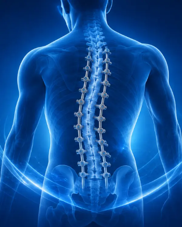
Spinal Fixation with Dr. Ahmed Sameh Nada
Some patients require Spinal Fusion and Instrumentation with screws and rods.
Possible indications include:
- Spondylolisthesis.
- Spinal instability.
- Selected spinal fractures.
- Certain deformities.
- Instability after extensive decompression.
- Selected spinal tumors.
Spondylolisthesis
Spondylolisthesis occurs when one vertebra slips relative to another.
Symptoms may include:
- Back pain.
- Leg pain.
- Nerve compression.
- Spinal instability.
Treatment depends on the degree of slippage, symptoms, and neurological findings.
Spinal Fractures
Spinal Fractures may occur because of:
- Trauma.
- Falls.
- Osteoporosis.
- Spinal tumors.
Treatment ranges from conservative management to stabilization surgery depending on spinal stability and neurological involvement.
Minimally Invasive Spine Surgery
Minimally Invasive Spine Surgery may be appropriate for selected conditions.
Techniques may include:
- Microdiscectomy.
- Minimally invasive fixation.
- Decompression.
- Endoscopic spinal procedures.
The appropriate technique depends on the underlying spinal condition.
Radiofrequency Treatment for Spine Pain
Radiofrequency Ablation, RFA, may be used for selected chronic spinal pain conditions.
It can be considered in some cases of:
- Facet joint pain.
- Chronic low back pain.
- Chronic neck pain related to facet joints.
- Sacroiliac joint pain.
The pain source should be identified before the procedure.
Does Radiofrequency Treat a Slipped Disc?
Not necessarily.
Radiofrequency is used for selected pain generators and does not remove a herniated disc that is compressing a nerve.
The cause of pain should therefore be confirmed before choosing treatment.
Traumatic Brain Injury with Dr. Ahmed Sameh Nada
The expertise of Dr. Ahmed Sameh Nada includes Traumatic Brain Injury and Emergency Neurosurgery.
This may involve:
- Acute intracranial hemorrhage.
- Epidural hematoma.
- Subdural hematoma.
- Skull fractures.
- Traumatic brain injury.
- Raised intracranial pressure.
- Selected complications after head trauma.
Epidural Hematoma
An Epidural Hematoma is a collection of blood between the skull and the outer covering of the brain.
It can occur after head trauma, and some cases require urgent surgery to relieve pressure on the brain.
Subdural Hematoma
A Subdural Hematoma is bleeding between the layers covering the brain.
It may be:
- Acute after trauma.
- Chronic, particularly in older adults.
Treatment depends on hematoma size, symptoms, and the degree of brain compression.
Hydrocephalus
Hydrocephalus is abnormal accumulation of cerebrospinal fluid within the brain’s ventricles.
Symptoms may include:
- Headache.
- Vomiting.
- Reduced consciousness.
- Walking difficulty.
- Different symptoms according to patient age and underlying cause.
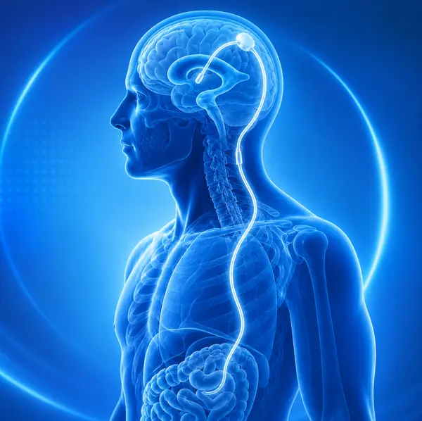
VP Shunt Surgery
In selected hydrocephalus cases, a Ventriculoperitoneal Shunt, VP Shunt, can be used to divert excess cerebrospinal fluid from the brain to the abdominal cavity.
The need for a shunt depends on the type and cause of hydrocephalus.
Dr. Ahmed Sameh Nada’s Treatment Approach
Dr. Ahmed Sameh Nada evaluates each neurological case comprehensively before recommending treatment.
Assessment may include:
- Neurological examination.
- CT and MRI review.
- CT Angiography or MR Angiography.
- Review of previous cerebral angiography.
- Evaluation of the relationship between lesions, nerves, and blood vessels.
- Comparison between endovascular and surgical options.
- Selection of the least invasive appropriate treatment.
- Structured follow-up after intervention.
Combining Neurosurgery and Neurointervention
One of the important aspects of Dr. Ahmed Sameh Nada’s practice is the combination of Neurosurgery and Neurointervention.
This is particularly valuable in cerebrovascular disease because some patients are best treated through catheter-based intervention, others through microsurgery, and selected complex cases may require a combined treatment strategy.
Dr. Ahmed Sameh Nada’s Clinical Experience
Dr. Ahmed Sameh Nada is a Consultant and Lecturer of Neurosurgery with expertise in brain surgery, spine surgery, cerebral and spinal vascular catheterization, and neurointervention.
His clinical practice includes complex neurovascular disease, brain and skull base tumors, spinal disorders, and emergency neurosurgical conditions.
Why Do Patients Consult Dr. Ahmed Sameh Nada?
Patients may seek an evaluation with Dr. Ahmed Sameh Nada for specialized management of:
Neurosurgery – neurointervention – cerebral angiography – brain aneurysm – aneurysm coiling – flow diverter – mechanical thrombectomy – stroke thrombectomy – AVM – DAVF – CCF – carotid artery stenting – brain tumor surgery – skull base surgery – pituitary surgery – lumbar disc surgery – cervical disc surgery – spinal fixation – minimally invasive spine surgery.
Book an Appointment with Dr. Ahmed Sameh Nada Through Macrocare
If you are searching for:
Dr. Ahmed Sameh Nada neurosurgeon
Dr. Ahmed Sameh Nada neurointervention
Dr. Ahmed Sameh Nada brain aneurysm
Dr. Ahmed Sameh Nada stroke thrombectomy
Dr. Ahmed Sameh Nada AVM
Dr. Ahmed Sameh Nada spine surgeon
Brain aneurysm specialist in Egypt
Neurointerventional surgeon in Egypt
Stroke thrombectomy specialist in Egypt
Spine surgeon in Egypt
or you need a specialist opinion regarding a brain aneurysm, acute stroke, cerebral AVM, DAVF, carotid stenosis, brain or skull base tumor, cervical disc disease, lumbar disc herniation, or another complex neurological condition, you can submit your case to Macrocare.
For an initial case review, patients should preferably provide:
- Medical reports.
- CT Brain.
- MRI Brain.
- CT Angiography.
- MR Angiography.
- Original cerebral angiography images or CD if previously performed.
- CT Perfusion or MR Perfusion in stroke cases if available.
- MRI Spine for spinal conditions.
- Previous operative reports.
- Pathology reports for tumor cases.
- Current medication list.
- Antiplatelet or anticoagulant medication list if relevant.
For Brain Aneurysm, AVM, DAVF, and other complex neurovascular conditions, the original angiography images are particularly valuable because vascular anatomy cannot always be fully assessed from the written report alone.
The Macrocare team can help organize the medical file and coordinate the appropriate consultation or appointment with Dr. Ahmed Sameh Nada.
Frequently Asked Questions About Dr. Ahmed Sameh Nada
What Is Dr. Ahmed Sameh Nada’s Specialty?
Dr. Ahmed Sameh Nada is a Consultant and Lecturer of Neurosurgery specializing in neurosurgery, neurointervention, cerebrovascular surgery, brain surgery, and spine surgery.
Is Dr. Ahmed Sameh Nada a Neurointerventional Specialist?
Yes. His practice includes cerebral and spinal vascular catheterization and neurointerventional procedures, including treatment of selected aneurysms and vascular malformations.
Does Dr. Ahmed Sameh Nada Treat Brain Aneurysms?
Yes. The expertise of Dr. Ahmed Sameh Nada includes assessment and treatment of Brain Aneurysms using endovascular or microsurgical techniques depending on aneurysm anatomy.
Can a Brain Aneurysm Be Treated Without Open Surgery?
In many patients, brain aneurysms can be treated using techniques such as Coiling, Stent-Assisted Coiling, or Flow Diversion. Other aneurysms are better treated using microsurgical clipping.
Does Dr. Ahmed Sameh Nada Perform Mechanical Thrombectomy for Stroke?
His neurointerventional practice includes Mechanical Thrombectomy for selected patients with acute large-vessel ischemic stroke when imaging and clinical criteria support intervention.
Does Dr. Ahmed Sameh Nada Treat Brain AVMs?
Yes. His expertise includes evaluation and treatment of Arteriovenous Malformations, AVM, using embolization, microsurgery, or multidisciplinary treatment depending on the case.
Does Dr. Ahmed Sameh Nada Treat DAVF?
Yes. Dural Arteriovenous Fistulas, DAVF, can be evaluated for endovascular or surgical treatment depending on their vascular anatomy and venous drainage.
Does Dr. Ahmed Sameh Nada Treat Carotid Artery Stenosis?
Yes. His neurovascular practice includes evaluation of carotid artery disease and determining whether medical therapy, carotid stenting, or another treatment is appropriate.
Does Dr. Ahmed Sameh Nada Perform Brain Tumor Surgery?
Yes. His expertise includes brain and skull base tumor surgery, including selected meningiomas, gliomas, pituitary tumors, acoustic neuromas, and metastatic brain tumors.
Does Dr. Ahmed Sameh Nada Treat Pituitary Tumors?
Yes. Selected pituitary tumors can be treated surgically, including by Endoscopic Transsphenoidal Surgery when appropriate.
Is Dr. Ahmed Sameh Nada a Spine Surgeon?
Yes. His neurosurgical practice includes spine surgery, including lumbar and cervical disc disease, spinal stenosis, spinal fixation, spondylolisthesis, and selected spinal fractures.
Does Every Slipped Disc Require Surgery?
No. Many disc herniations improve with conservative treatment. Surgery is usually considered when there is significant nerve compression, progressive weakness, persistent symptoms, or urgent neurological warning signs.
Does Dr. Ahmed Sameh Nada Use Radiofrequency for Spine Pain?
Radiofrequency procedures may be used for selected chronic spinal pain conditions after confirming the source of pain.
What Imaging Should I Send for a Brain Aneurysm or AVM Review?
It is useful to send CT or MRI, CT Angiography or MR Angiography, and the original cerebral angiography images if an angiogram has already been performed.
What Imaging Should I Send for a Spine Surgery Review?
An MRI of the affected spinal region is usually one of the most important studies, together with any CT scans, previous X-rays, operative reports, and treatment history.
How Can I Book an Appointment with Dr. Ahmed Sameh Nada?
You can contact Macrocare and submit the patient’s reports and imaging. The Macrocare team can help organize the medical file and coordinate the appropriate consultation or appointment with Dr. Ahmed Sameh Nada.