Genel Bakış
Misr Radiology Center (MRC) – Advanced Diagnostic and Interventional Radiology Center in Egypt
Misr Radiology Center (MRC) is one of Egypt’s established centers for advanced diagnostic radiology, nuclear medicine, molecular imaging, interventional radiology, theranostics, women’s imaging, and specialized medical imaging.
Founded in 1989, Misr Radiology Center has developed into a multidisciplinary imaging network providing diagnostic and image-guided therapeutic services through specialized radiologists, nuclear medicine physicians, interventional radiologists, technicians, and supporting medical teams.
MRC provides advanced imaging services including MRI, 3 Tesla MRI, PET/MRI, PET/CT, CT Scan, Digital X-Ray, Ultrasound, Doppler, Mammography, DEXA, Nuclear Medicine, Dental CBCT, EEG, EMG, image-guided biopsy, interventional radiology, and targeted radionuclide therapy.
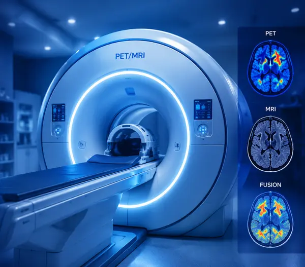
One of the center’s most distinctive technologies is PET/MRI, which MRC introduced as the first installation of its kind in Africa and the Middle East according to the center. Its New Cairo facility combines PET molecular imaging with advanced 3 Tesla MRI in a single examination.
Through Macrocare, local and international patients can request appointments, submit medical reports, arrange advanced radiology examinations, and coordinate imaging or interventional radiology services at Misr Radiology Center in Egypt.
Misr Radiology Center Services
Services available across the MRC network include:
- MRI – Magnetic Resonance Imaging.
- 1.5 Tesla MRI.
- 3 Tesla MRI.
- Functional MRI – fMRI.
- MR Spectroscopy.
- MR Perfusion.
- MR Diffusion.
- MR Tractography.
- Multiparametric MRI.
- PET/MRI.
- FDG PET/MRI.
- FDOPA PET/MRI.
- PSMA PET/MRI.
- DOTATATE PET/MRI.
- PET/CT.
- FDG PET/CT.
- Gallium PSMA PET/CT.
- Gallium DOTATATE PET/CT.
- CT Scan.
- CT Angiography.
- Digital X-Ray.
- Fluoroscopy.
- Ultrasound.
- Color Doppler.
- Mammography.
- Breast MRI.
- Breast Ultrasound.
- Image-guided breast biopsy.
- DEXA Scan.
- Body Composition Analysis.
- Nuclear Medicine.
- Theranostics.
- Lutetium-177 PSMA Therapy.
- Lutetium-177 DOTATATE Therapy.
- Interventional Radiology.
- Image-guided biopsies.
- Liver tumor ablation.
- TACE.
- DEB-TACE.
- Y-90 Radioembolization.
- Uterine Fibroid Embolization.
- Prostatic Artery Embolization.
- Varicocele Embolization.
- Vertebroplasty.
- Venous access procedures.
- Drainage procedures.
- Dental CBCT.
- TMJ imaging.
- EEG.
- EMG.
PET/MRI at Misr Radiology Center
PET/MRI is one of the most advanced hybrid imaging technologies available at Misr Radiology Center.
The examination combines:
- The metabolic and molecular information of PET.
- High-resolution anatomical MRI.
- Diffusion imaging.
- Perfusion imaging.
- Spectroscopy when required.
- Functional and multiparametric MRI techniques.
MRC states that its PET/MRI system was the first PET/MRI installation in Africa and the Middle East.
What Is PET/MRI?
Positron Emission Tomography / Magnetic Resonance Imaging – PET/MRI combines two complementary imaging technologies.
PET evaluates biological and metabolic activity after administration of an appropriate radioactive tracer.
MRI provides highly detailed anatomical and functional information without using the ionizing radiation associated with CT imaging.
The combination can be especially useful when both molecular activity and detailed soft-tissue characterization are important.
PET/MRI for Cancer
PET/MRI can be used in selected oncology cases for:
- Tumor detection.
- Local tumor staging.
- Whole-body assessment.
- Treatment response evaluation.
- Detection of recurrence.
- Assessment of metastatic disease.
- Differentiating viable tumor from treatment-related changes.
MRC uses PET/MRI for a range of oncological and neurological applications.
PET/MRI FDG Oncology
FDG PET/MRI combines FDG metabolic imaging with high-resolution MRI.
It can be considered in selected cancers where detailed local MRI information and whole-body metabolic assessment are both clinically useful.
Applications may include selected:
- Breast cancers.
- Rectal cancers.
- Gynecologic tumors.
- Head and neck cancers.
- Liver tumors.
- Pancreatic tumors.
- Renal tumors.
- Pediatric malignancies.
- Multiple myeloma assessment.
PET/MRI FDG Brain
Brain FDG PET/MRI combines metabolic brain imaging with detailed structural MRI.
It may be used in selected cases involving:
- Epilepsy.
- Brain tumors.
- Neurodegenerative disorders.
- Dementia evaluation.
- Selected inflammatory neurological disorders.
PET/MRI FDOPA Brain
FDOPA PET/MRI – Brain is an advanced molecular imaging examination particularly relevant in selected neurological and neuro-oncology cases.
FDOPA imaging may be useful in the assessment of:
- Selected brain tumors.
- Gliomas.
- Post-treatment brain tumor evaluation.
- Parkinsonian disorders in selected circumstances.
MRC lists FDOPA PET/MRI Brain among its international patient imaging services.
PSMA PET/MRI for Prostate Cancer
Gallium PSMA PET/MRI combines prostate-specific molecular imaging with high-resolution MRI.
It may be used in selected patients for:
- Prostate cancer staging.
- Assessment of suspected recurrence.
- Detection of metastatic disease.
- Detailed evaluation of pelvic disease.
- Treatment planning.
MRC specifically lists Gallium PSMA PET/MRI for prostate cancer among its PET/MRI examinations.
DOTATATE PET/MRI
Gallium DOTATATE PET/MRI is used for molecular imaging of selected neuroendocrine tumors.
The examination combines somatostatin-receptor-targeted PET imaging with detailed MRI information.
It may assist in:
- Tumor localization.
- Staging.
- Metastatic assessment.
- Evaluation before targeted radionuclide therapy.
PET/CT at Misr Radiology Center
PET/CT combines metabolic PET imaging with CT-based anatomical localization.
It is widely used in oncology for:
- Cancer staging.
- Restaging.
- Treatment-response evaluation.
- Detection of recurrence.
- Metastatic assessment.
- Selection of an appropriate biopsy target in selected patients.
MRC provides conventional FDG PET/CT as well as advanced Gallium-based studies.
FDG PET/CT
FDG PET/CT evaluates glucose metabolism within tissues.
It is frequently used in the assessment of various cancers and may also have selected neurological, inflammatory, and infectious applications.
Gallium PSMA PET/CT
PSMA PET/CT is an important molecular imaging technique for prostate cancer.
It can be used in selected patients for:
- Primary staging.
- Biochemical recurrence.
- Assessment of lymph nodes.
- Bone metastases.
- Distant metastatic disease.
- Selection of patients for PSMA-targeted therapy.
MRC identifies itself as an early provider in Egypt of advanced Gallium PET/CT examinations for prostate and neuroendocrine tumors.
Gallium DOTATATE PET/CT
Gallium DOTATATE PET/CT targets somatostatin receptors and is particularly useful in selected neuroendocrine tumors.
It may be used for:
- Primary tumor localization.
- Staging.
- Metastatic assessment.
- Evaluation of receptor expression.
- Selection of patients for PRRT.
PET/MRI vs PET/CT
The most appropriate examination depends on the disease and clinical question.
PET/MRI can offer particularly detailed soft-tissue and functional MRI information while reducing CT-related radiation exposure.
PET/CT remains highly valuable and may be preferred for other indications, including many conventional oncology staging and follow-up pathways.
MRC provides both technologies, allowing the examination to be selected according to the individual case.
MRI at Misr Radiology Center
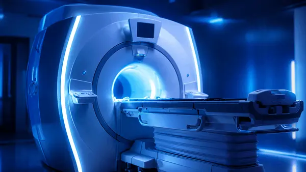
Magnetic Resonance Imaging – MRI uses magnetic fields and radiofrequency signals to produce detailed images without ionizing radiation.
MRC provides MRI services including 1.5 Tesla and advanced 3 Tesla imaging, together with specialized metabolic and functional MRI techniques.
3 Tesla MRI
3T MRI provides a strong magnetic field that can support high-resolution and advanced functional imaging protocols.
MRC uses 3 Tesla MRI for advanced applications involving:
- Brain.
- Spine.
- Musculoskeletal imaging.
- Abdomen.
- Pelvis.
- Oncology.
- Prostate.
- Neurological assessment.
- Functional imaging.
Brain MRI
Brain MRI may be requested for:
- Brain tumors.
- Stroke.
- Epilepsy.
- Multiple sclerosis.
- Headaches with neurological indications.
- Pituitary disease.
- Cranial nerve disorders.
- Neurodegenerative disease.
- Preoperative planning.
Spine MRI
MRI is frequently used to evaluate:
- Cervical disc disease.
- Lumbar disc herniation.
- Spinal stenosis.
- Spinal cord compression.
- Spinal tumors.
- Infection.
- Inflammatory disease.
- Postoperative conditions.
MR Spectroscopy
MR Spectroscopy evaluates the biochemical composition of tissue rather than anatomy alone.
MRC performs advanced MR spectroscopy protocols, particularly for neurological and brain-lesion assessment.
It can be useful in selected cases for:
- Brain tumor characterization.
- Differentiating tumor from non-neoplastic abnormalities.
- Treatment follow-up.
- Metabolic assessment of brain lesions.
MR Perfusion
MR Perfusion assesses blood flow and vascular characteristics within tissue.
MRC performs several perfusion techniques, including contrast and non-contrast approaches.
Potential applications include:
- Brain tumors.
- Stroke.
- Epilepsy.
- Selected neurological disorders.
MR Diffusion
Diffusion MRI evaluates movement of water molecules within tissue.
It is particularly important in:
- Acute stroke.
- Brain tumors.
- Infection.
- Oncological imaging.
- Tissue characterization.
MR Tractography
MR Tractography maps major white-matter pathways in the brain and spinal cord.
It can be valuable for:
- Brain tumor surgery planning.
- Neurosurgical assessment.
- Evaluation of lesions near important neural pathways.
Functional MRI – fMRI
Functional MRI – fMRI maps brain activity by identifying changes associated with neurological function.
MRC performs functional MRI protocols for functions including:
- Motor activity.
- Language.
- Vision.
- Auditory function.
- Memory.
It can be particularly valuable before surgery for brain tumors, epilepsy, and selected vascular malformations.
Multiparametric MRI
Multiparametric MRI combines several MRI sequences to improve tissue characterization.
An important application is multiparametric prostate MRI, which can help evaluate suspected prostate cancer and guide further assessment.
CT Scan at Misr Radiology Center
Computed Tomography – CT Scan uses X-rays and computer processing to produce detailed cross-sectional images.
CT may be requested for:
- Trauma.
- Chest disease.
- Lung conditions.
- Abdominal disease.
- Kidney and urinary tract conditions.
- Internal bleeding.
- Tumor evaluation.
- Vascular disease.
- Emergency imaging.
CT Angiography
CT Angiography – CTA evaluates arteries and veins after administration of contrast material.
Applications include assessment of:
- Cerebral arteries.
- Carotid arteries.
- Aorta.
- Pulmonary arteries.
- Peripheral arteries.
- Coronary arteries in selected examinations.
Digital X-Ray
MRC provides Digital X-Ray for a broad range of examinations including:
- Bone fractures.
- Chest imaging.
- Joint assessment.
- Spine imaging.
- Selected abdominal studies.
Digital X-ray is part of MRC’s diagnostic radiology service portfolio.
Fluoroscopy
Fluoroscopy uses continuous or real-time X-ray imaging to visualize movement and passage of contrast material.
It may be used for selected:
- Gastrointestinal examinations.
- Contrast studies.
- Swallowing studies.
- Urinary tract examinations.
- Image-guided procedures.
Ultrasound at Misr Radiology Center
Ultrasound uses high-frequency sound waves to produce images without ionizing radiation.
Applications include:
- Abdominal ultrasound.
- Liver imaging.
- Kidney ultrasound.
- Thyroid ultrasound.
- Pelvic ultrasound.
- Pregnancy imaging.
- Breast ultrasound.
- Soft-tissue imaging.
Doppler Ultrasound
Color Doppler and Duplex Ultrasound evaluate blood flow within arteries and veins.
They may be used to assess:
- Carotid arteries.
- Peripheral arteries.
- Venous thrombosis.
- Varicose veins.
- Testicular blood flow.
- Renal vessels.
- Other vascular conditions.
Women’s Health Unit at Misr Radiology Center
MRC operates a dedicated Women’s Health Unit combining several diagnostic services, including breast imaging, DEXA, pregnancy imaging, and pelvic imaging.
Mammography at MRC
Digital Mammography is used for breast cancer screening and diagnostic breast assessment.
MRC’s breast imaging services include:
- Digital Mammogram.
- Breast MRI.
- Breast Ultrasound.
- Ultrasound-guided biopsy.
- Stereotactic breast biopsy.
- Needle localization.
- Cyst aspiration.
- Fine Needle Aspiration.
Breast MRI
Breast MRI may be used in selected patients for:
- High-risk screening.
- Extent-of-disease assessment.
- Implant assessment.
- Evaluation of specific mammographic or ultrasound findings.
- Treatment monitoring.
Breast Ultrasound
Breast ultrasound may help evaluate:
- Breast masses.
- Cysts.
- Dense breast tissue.
- Findings identified by mammography.
- Image-guided biopsy targets.
Image-Guided Breast Biopsy
When imaging identifies a suspicious breast abnormality, tissue sampling may be required.
MRC provides techniques including:
- Ultrasound-guided breast biopsy.
- Stereotactic-guided biopsy.
- Fine Needle Aspiration.
- Localization procedures.
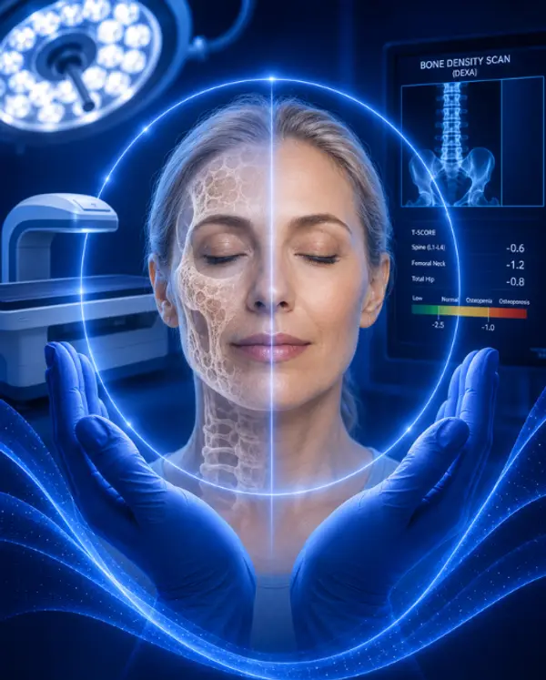
DEXA Scan
DEXA – Dual-Energy X-Ray Absorptiometry measures bone mineral density.
It is commonly used to diagnose and monitor:
- Osteoporosis.
- Osteopenia.
- Bone-density changes.
Body Composition Analysis
MRC also uses DEXA technology for Body Composition Analysis, including assessment of:
- Body fat.
- Lean muscle mass.
- Bone composition.
This may be useful in obesity management, clinical nutrition, rehabilitation, and athletic assessment.
Nuclear Medicine at Misr Radiology Center
Nuclear Medicine uses small quantities of radioactive tracers to evaluate organ function and disease activity.
MRC provides nuclear medicine and molecular imaging as part of its diagnostic and therapeutic services.
Applications may include:
- Bone scanning.
- Thyroid assessment.
- Renal function studies.
- Selected cardiac studies.
- Oncology imaging.
Theranostics at Misr Radiology Center
Theranostics combines targeted molecular diagnosis with targeted radionuclide therapy.
The principle is to identify whether cancer cells express a specific molecular target and, when appropriate, use a radioactive therapeutic agent directed at the same target.
MRC provides targeted Lutetium therapies for prostate cancer and neuroendocrine tumors.
Lutetium-177 PSMA Therapy
177Lu-PSMA Therapy is a targeted radionuclide treatment used in selected patients with advanced PSMA-positive prostate cancer.
Eligibility typically depends on factors such as:
- Disease stage.
- Previous treatments.
- PSMA imaging.
- Blood tests.
- Kidney function.
- Bone marrow reserve.
- Overall clinical condition.
Lutetium-177 DOTATATE Therapy
177Lu-DOTATATE, also known as Peptide Receptor Radionuclide Therapy – PRRT, is used for selected somatostatin-receptor-positive neuroendocrine tumors.
Patients generally require appropriate receptor imaging before treatment.
Interventional Radiology at Misr Radiology Center
Interventional Radiology uses imaging such as ultrasound, CT, fluoroscopy, and angiography to guide minimally invasive diagnostic and therapeutic procedures.
MRC has a dedicated interventional radiology service and uses hybrid CT/angiography guidance for a range of procedures.
Liver Tumor Treatment
MRC’s interventional radiology services include several approaches for selected liver tumors.
These include:
- Radiofrequency Ablation.
- Microwave Ablation.
- TACE.
- DEB-TACE.
- Y-90 Radioembolization.
Radiofrequency Ablation
Radiofrequency Ablation – RFA uses thermal energy delivered through an image-guided probe to destroy selected tumor tissue.
It can be considered for certain:
- Liver tumors.
- Bone tumors.
- Other selected lesions.
Microwave Ablation
Microwave Ablation is another thermal ablation technique used to treat selected tumors under imaging guidance.
The most suitable technique depends on tumor size, location, anatomy, and surrounding structures.
TACE for Liver Cancer
Transarterial Chemoembolization – TACE is used in selected liver cancers, particularly hepatocellular carcinoma.
A catheter is advanced into arteries supplying the tumor, allowing targeted delivery of therapy and embolization of tumor blood flow.
MRC lists conventional TACE and DEB-TACE among its interventional oncology procedures.
Y-90 Radioembolization
Yttrium-90 Radioembolization is a catheter-based liver-directed treatment using radioactive microspheres.
MRC lists Y-90 transarterial radioembolization among its interventional liver procedures.
Uterine Fibroid Embolization
Uterine Fibroid Embolization – UFE is a minimally invasive treatment for selected symptomatic uterine fibroids.
The procedure blocks blood flow to the fibroids through uterine arteries, causing them to shrink over time.
MRC includes uterine fibroid embolization among its interventional radiology treatments.
Prostatic Artery Embolization
Prostatic Artery Embolization – PAE is an image-guided treatment for selected men with Benign Prostatic Hyperplasia – BPH.
The procedure reduces blood supply to parts of the enlarged prostate with the aim of reducing its volume and urinary symptoms.
MRC provides PAE within its interventional radiology portfolio.
Varicocele Embolization
Varicocele Embolization is a catheter-based treatment for selected varicoceles.
It aims to block abnormal veins responsible for venous reflux without conventional open surgery.
Vertebroplasty
Vertebroplasty is an image-guided procedure in which bone cement is injected into selected vertebral compression fractures.
It may be considered in carefully selected patients with painful fractures.
Image-Guided Biopsy at Misr Radiology Center
MRC offers CT-guided and ultrasound-guided biopsies for multiple body sites.
These include selected biopsies involving:
- Liver.
- Kidney.
- Pancreas.
- Spleen.
- Lymph nodes.
- Lung.
- Bone.
- Thyroid.
- Breast.
- Abdominal masses.
Port-A-Cath and Venous Access
MRC’s interventional services also include venous access procedures such as:
- Port-A-Cath insertion.
- PICC Line placement.
- Central venous catheter placement.
- Permcath insertion.
Nephrostomy and Urinary Interventions
Interventional radiology services include selected urinary drainage procedures such as:
- Percutaneous nephrostomy.
- Percutaneous ureteric stent placement.
These may be required in patients with urinary obstruction.
Drainage of Collections and Abscesses
Image-guided drainage can be used for selected:
- Abscesses.
- Abdominal collections.
- Pleural fluid.
- Ascites.
- Other fluid collections.
Dental CBCT at Misr Radiology Center
MRC operates a specialized Dental and Maxillofacial Imaging Unit offering panoramic imaging and advanced Cone Beam CT – CBCT.
Services include:
- Dental implant planning.
- Single-tooth CBCT.
- Single-quadrant CBCT.
- Single-arch imaging.
- Both-arch imaging.
- Full-face CBCT.
- Endodontic high-resolution imaging.
- Cephalometric analysis.
- Orthodontic imaging.
- ENT imaging.
- Temporal bone imaging.
- Cochlear implant protocol.
- TMJ analysis.
- 4D TMJ imaging.
- Panoramic X-ray.
CBCT for Dental Implants
CBCT provides three-dimensional visualization of the jaws and surrounding anatomy.
It can help with:
- Dental implant planning.
- Bone-volume assessment.
- Assessment of the mandibular canal.
- Sinus evaluation.
- Surgical planning.
TMJ Imaging
MRC’s dental imaging unit provides TMJ analysis, including advanced dynamic 4D TMJ imaging capabilities.
EEG at Misr Radiology Center
MRC also offers Electroencephalography – EEG among its non-radiology diagnostic services.
EEG evaluates electrical brain activity and is commonly used in the assessment of:
- Epilepsy.
- Seizures.
- Selected disorders of consciousness.
- Other neurological conditions.
EMG at Misr Radiology Center
Electromyography – EMG evaluates muscle and peripheral nerve function.
It may be requested in patients with:
- Numbness.
- Weakness.
- Peripheral nerve injury.
- Nerve compression.
- Neuromuscular disease.
Anesthesia Support for Imaging and Procedures
MRC states that anesthesia support is available for patients undergoing examinations or procedures that require sedation or anesthesia. This can be particularly relevant for selected MRI, PET/MRI, pediatric, interventional, or claustrophobic patients.
Misr Radiology Center for International Patients
MRC has a dedicated Medical Tourism offering for international patients requiring advanced imaging or interventional procedures in Egypt.
International services described by the center include:
- Advanced imaging coordination.
- PET/MRI.
- PET/CT.
- Theranostics.
- Interventional radiology.
- Advanced biopsy.
- 3T MRI.
- Screening programs.
- Accommodation coordination.
- Airport meet-and-assist.
- Transportation.
- Tourism arrangements.
Through Macrocare, international patients can also coordinate their imaging appointment together with the wider medical journey in Egypt.
Why International Patients Choose Misr Radiology Center
MRC can be particularly relevant to international patients looking for access in Egypt to:
- PET/MRI.
- FDOPA Brain PET/MRI.
- PSMA PET imaging.
- DOTATATE PET imaging.
- Advanced 3T MRI.
- Functional MRI.
- Interventional oncology.
- Lutetium therapy.
- Advanced image-guided biopsy.
- Minimally invasive interventional radiology procedures.
Misr Radiology Center Branches
MRC currently lists locations across Cairo including:
- New Cairo / Fifth Settlement: Off 90 Street, near MRC Square, between Air Force Specialized Hospital and Lake View Compound.
- Heliopolis – Khalifa El Maamoun: 8 Khalifa El Maamoun Street.
- Heliopolis – El Merghany: 11 El Merghany Street.
- Nasr City – Misr Scan: 8 Mostafa El Nahas Street.
MRI 24 Hours at Selected MRC Branches
MRC states that the MRI department operates 24 hours at the Khalifa El Maamoun and Nasr City branches, while general working hours vary by location and day.
Why Choose Misr Radiology Center?
Patients may consider Misr Radiology Center because it combines a broad spectrum of advanced imaging and minimally invasive treatment services within one radiology network.
Key capabilities include:
- Established in 1989.
- Advanced diagnostic radiology.
- Nuclear medicine.
- PET/CT.
- PET/MRI.
- First PET/MRI installation in Africa and the Middle East according to MRC.
- Advanced 3 Tesla MRI.
- Functional and metabolic MRI.
- Dedicated interventional radiology services.
- Theranostics and Lutetium therapy.
- Women’s imaging.
- Advanced dental CBCT.
- Multiple branches across Cairo.
- International patient services.
Book Misr Radiology Center Through Macrocare
If you are searching for:
Misr Radiology Center
MRC Egypt
Misr Radiology Center Egypt
Misr Radiology Center Cairo
Misr Radiology Center New Cairo
Misr Radiology Center Heliopolis
Misr Radiology Center Nasr City
Misr Radiology Center PET MRI
PET MRI Egypt
PET MRI Cairo
FDOPA PET MRI Egypt
PSMA PET MRI Egypt
Gallium PSMA PET CT Egypt
DOTATATE PET CT Egypt
3 Tesla MRI Egypt
Functional MRI Egypt
Interventional Radiology Egypt
Lutetium Therapy Egypt
Lutetium PSMA Egypt
Lutetium DOTATATE Egypt
Prostatic Artery Embolization Egypt
Uterine Fibroid Embolization Egypt
Liver Tumor Ablation Egypt
TACE Egypt
Y90 Radioembolization Egypt
you can submit the requested examination or medical case to Macrocare.
Macrocare can assist with:
- Identifying the required examination.
- Reviewing the physician’s radiology request.
- Coordinating the MRC appointment.
- Confirming preparation requirements.
- Coordinating contrast or anesthesia requirements when applicable.
- Organizing international patient logistics.
- Coordinating treatment when an interventional procedure is required.
What Should I Send Before Booking an Imaging Examination?
Patients should preferably provide:
- Physician request.
- Diagnosis.
- Previous imaging reports.
- Original CT/MRI/PET images when relevant.
- Pathology report for oncology patients.
- Current treatment history.
- Previous surgery reports.
- Kidney function tests when contrast may be required.
- Medication list.
- Pregnancy status when relevant.
- Any history of contrast allergy.
- Pacemaker or implanted-device details before MRI.
What Should Oncology Patients Send?
For oncology imaging such as PET/CT, PET/MRI, PSMA, DOTATATE, or advanced MRI, it is useful to provide:
- Pathology report.
- Cancer diagnosis and stage.
- Previous PET imaging.
- CT and MRI.
- Surgical reports.
- Chemotherapy history.
- Radiotherapy history.
- Current systemic treatment.
- Tumor markers when relevant.
What Should Patients Send for Interventional Radiology?
For interventional radiology procedures, send:
- Current diagnosis.
- Recent imaging.
- Laboratory tests.
- CBC.
- Coagulation profile when available.
- Kidney and liver function according to procedure.
- Current medications.
- Anticoagulant or antiplatelet medications.
- Previous treatment reports.
Frequently Asked Questions About Misr Radiology Center
What Is Misr Radiology Center?
Misr Radiology Center – MRC is an advanced diagnostic and interventional radiology center in Egypt providing MRI, CT, PET/CT, PET/MRI, nuclear medicine, interventional radiology, women’s imaging, dental imaging, and other specialized services.
When Was Misr Radiology Center Established?
Misr Radiology Center was established in 1989.
Does Misr Radiology Center Have PET/MRI?
Yes. MRC provides PET/MRI at its New Cairo facility and states that it introduced the first PET/MRI system in Africa and the Middle East.
Does MRC Offer FDOPA PET/MRI Brain?
Yes. FDOPA PET/MRI Brain is listed among the PET/MRI examinations offered through MRC’s international patient program.
Does MRC Offer PSMA PET Scans?
Yes. MRC provides Gallium PSMA PET/CT and PSMA PET/MRI for selected prostate cancer patients.
Does Misr Radiology Center Offer DOTATATE PET?
Yes. MRC provides Gallium DOTATATE PET/CT and PET/MRI for selected neuroendocrine tumor patients.
Does MRC Provide Lutetium Therapy?
Yes. MRC provides targeted radionuclide therapies including Lutetium PSMA and Lutetium DOTATATE.
Does Misr Radiology Center Have 3 Tesla MRI?
Yes. MRC provides 3 Tesla MRI and advanced functional and metabolic MRI protocols.
Does MRC Perform Functional MRI?
Yes. MRC performs advanced Functional MRI – fMRI, including selected motor, language, visual, auditory, and memory mapping protocols.
Does MRC Perform MR Spectroscopy?
Yes. MR Spectroscopy is included among MRC’s advanced MRI capabilities.
Does MRC Perform Interventional Radiology?
Yes. MRC provides a broad range of Interventional Radiology procedures for vascular, oncological, urological, gynecological, and other conditions.
Does MRC Treat Liver Tumors with Interventional Radiology?
Yes. MRC lists procedures including radiofrequency ablation, microwave ablation, TACE, DEB-TACE and Y-90 radioembolization for selected liver tumors.
Does MRC Perform Uterine Fibroid Embolization?
Yes. Uterine Fibroid Embolization – UFE is included among MRC’s interventional radiology procedures.
Does MRC Perform Prostatic Artery Embolization?
Yes. Prostatic Artery Embolization – PAE for selected patients with benign prostate enlargement is included among MRC’s interventional services.
Does MRC Treat Varicocele by Embolization?
Yes. Varicocele Embolization is listed among MRC’s interventional radiology procedures.
Does MRC Perform Image-Guided Biopsies?
Yes. MRC provides CT- and ultrasound-guided biopsies for multiple organs including liver, kidney, lung, pancreas, lymph nodes, bone, breast, and thyroid.
Does Misr Radiology Center Offer Mammography?
Yes. MRC has a dedicated Women’s Health Unit providing digital mammography, breast ultrasound, breast MRI, and image-guided breast procedures.
Does MRC Provide DEXA Bone Density Scan?
Yes. DEXA is available for bone mineral density evaluation and body composition assessment.
Does Misr Radiology Center Provide Dental CBCT?
Yes. MRC has a specialized dental imaging unit offering CBCT, panoramic imaging, implant planning, orthodontic imaging, and TMJ assessment.
Where Are Misr Radiology Center Branches?
MRC lists branches in New Cairo, Heliopolis, and Nasr City, including the Misr Scan location.
Can International Patients Book Misr Radiology Center?
Yes. MRC has services directed at international patients, and Macrocare can coordinate the imaging examination or interventional procedure as part of the patient’s medical journey in Egypt.
How Can I Book Misr Radiology Center Through Macrocare?
Send the required examination, physician request, medical reports, and previous imaging to Macrocare. The team can coordinate the appropriate MRC service, appointment, required preparation, and international patient arrangements when needed.

Misr Radiology Center (MRC) tedavileri
Bu sağlayıcının sunduğu işlemler, gerçek başlangıç fiyatlarıyla.
Kemik Yoğunluğu Taraması ( DEXA )

MR Taraması

Biyopsi Analizi
Radyoterapi
En az 1 gün
PET MRI scan
PET/MRI FDOPA – Brain
Yalnızca belirtilen fiyatlar — uçuş ve konaklama ayrıca hesaplanır. Nihai maliyet ücretsiz teklifinizde onaylanır. 2026-07-20 tarihli kurlar.
Sunulan diğer hizmetler
Bu hizmetler bu sağlık kuruluşunda mevcuttur; fiyatlar talep üzerine bildirilir.